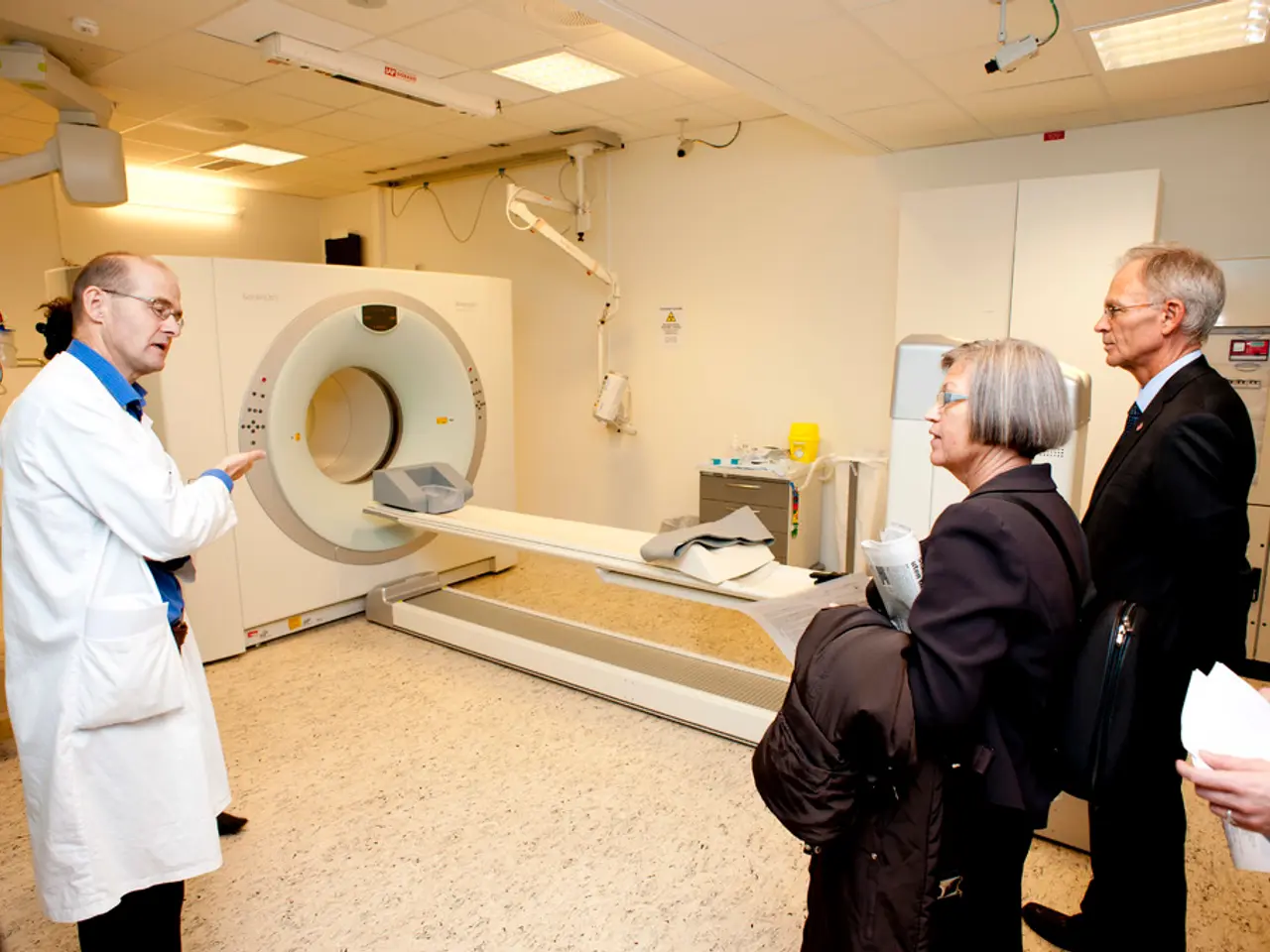
MRI of Ovarian Cancer: Potential Revealings
In the diagnosis and staging of ovarian cancer, a variety of tests are utilised to assess tumor presence, local and advanced disease spread, and tumor markers indicative of the disease.
One such test is the MRI scan, a minimally invasive outpatient procedure that typically takes 45-60 minutes to complete. Previous staging has taken place with techniques that do not affect fertility. It's worth noting that MRI scans are not typically used to check for ovarian cancer, but they can help with staging and examining areas to which the cancer may have spread.
Pelvic or transvaginal ultrasound (TVUS) is useful for early detection of ovarian tumors by imaging the ovaries locally. CT scans are used to detect advanced disease and evaluate abdominal involvement. PET-CT scans provide whole-body imaging to evaluate metastatic spread and help in precise staging. Blood tests measuring CA-125, a tumor marker often elevated in ovarian cancer, assist in diagnosis and monitoring recurrence. Biopsy, when performed, confirms the diagnosis histologically by sampling ovarian tissue. Genetic tests identify inherited mutations (such as in BRCA1/2), which have implications for risk assessment and treatment planning.
In some cases, an MRI with or without a contrast dye may be appropriate to assist doctors with determining the stage of ovarian cancer at the point of diagnosis or confirming a recurrence. A person will change into a hospital gown and receive a contrast dye injection before the MRI. During the procedure, the person will hear intermittent banging noises. Some facilities may offer items like hearing plugs to help keep the person comfortable during the MRI.
Doctors may use an MRI to get more information about areas that are difficult to see using a CT scan. The results of an MRI can help a doctor determine whether a surgeon will easily be able to remove the tumor. It's important to note that despite the safety of MRI scans, doctors typically avoid using them to examine the ovaries because a person's movements can interfere with the results more easily than they can with a CT scan. An MRI can show a detailed image of the uterus, lymph nodes, and other surrounding areas.
A laparoscopy involves a surgeon inserting a small tube with a light and camera into the abdomen to check for signs of ovarian cancer. A colonoscopy can check the large intestine for signs that the cancer has spread. Blood tests can check blood cell counts, liver and kidney function, and CA-125 levels, a marker for ovarian cancer.
In cases where a CT scan cannot show a small ovarian tumor but can help determine if the cancer has spread to nearby tissues or organs, an MRI may be recommended. Similarly, if the results of a CT scan are inconclusive, an MRI may be used to provide more detailed images.
It's crucial to remember that the comprehensive diagnostic pathway typically integrates imaging modalities (ultrasound, MRI, CT, PET-CT), serum tumor markers (CA-125), tissue biopsy, and genetic profiling to accurately diagnose and stage ovarian cancer.
An exploratory laparotomy involves a surgeon making an incision in the abdomen to examine the ovaries and other organs in the pelvis, and removing the ovaries, tumor mass, and uterus if cancer is present. This procedure is usually performed if the diagnosis is confirmed through other tests.
Emerging diagnostic approaches also include liquid biopsies (like the Avantect® test) and multi-cancer early detection blood tests, coupled with whole-body MRI for more sensitive screening and early detection.
[1] National Cancer Institute. (2021). Ovarian Cancer Staging. Retrieved from https://www.cancer.gov/types/ovarian/hp/ovarian-staging-pdq [2] American Cancer Society. (2021). Diagnosing Ovarian Cancer. Retrieved from https://www.cancer.org/cancer/ovarian-cancer/detection-diagnosis-staging/how-ovarian-cancer-is-diagnosed.html [3] American College of Radiology. (2020). Ovarian Cancer Screening. Retrieved from https://www.acr.org/-/media/ACR/Files/Radiology-Residency/Program-Directors/Resident-Resources/Ovarian-Cancer-Screening.pdf [4] Mayo Clinic. (2021). Ovarian cancer diagnosis. Retrieved from https://www.mayoclinic.org/diseases-conditions/ovarian-cancer/diagnosis-treatment/drc-20374903
- In the staging and examination of potential spread for ovarian cancer, medical professionals might utilize tests like MRI scans, pelvic or transvaginal ultrasound (TVUS), CT scans, PET-CT scans, and blood tests measuring CA-125 levels.
- Although MRI scans are not typically used for ovarian cancer screening, they can aid in the staging process, helping doctors determine if the cancer has spread and providing more detailed images of the uterus, lymph nodes, and surrounding areas.
- The comprehensive diagnostic pathway for ovarian cancer often integrates imaging modalities such as MRI, CT, and PET-CT, serum tumor markers like CA-125, tissue biopsy, and genetic profiling to accurately diagnose and stage the disease.
- In addition to traditional diagnostic methods, emerging approaches in health and wellness aim to improve early detection of ovarian cancer through liquid biopsies and multi-cancer early detection blood tests, combined with whole-body MRI for sensitive screening.


