Exploring and Foreseeing the Uses of PET Radiomics in Radiotherapy
In the realm of cancer care, groundbreaking advancements are being made at the intersection of Positron Emission Tomography (PET) imaging, radiomics, and artificial intelligence (AI). These innovations are transforming the way we diagnose, treat, and understand cancer, paving the way for a new era of personalized oncology.
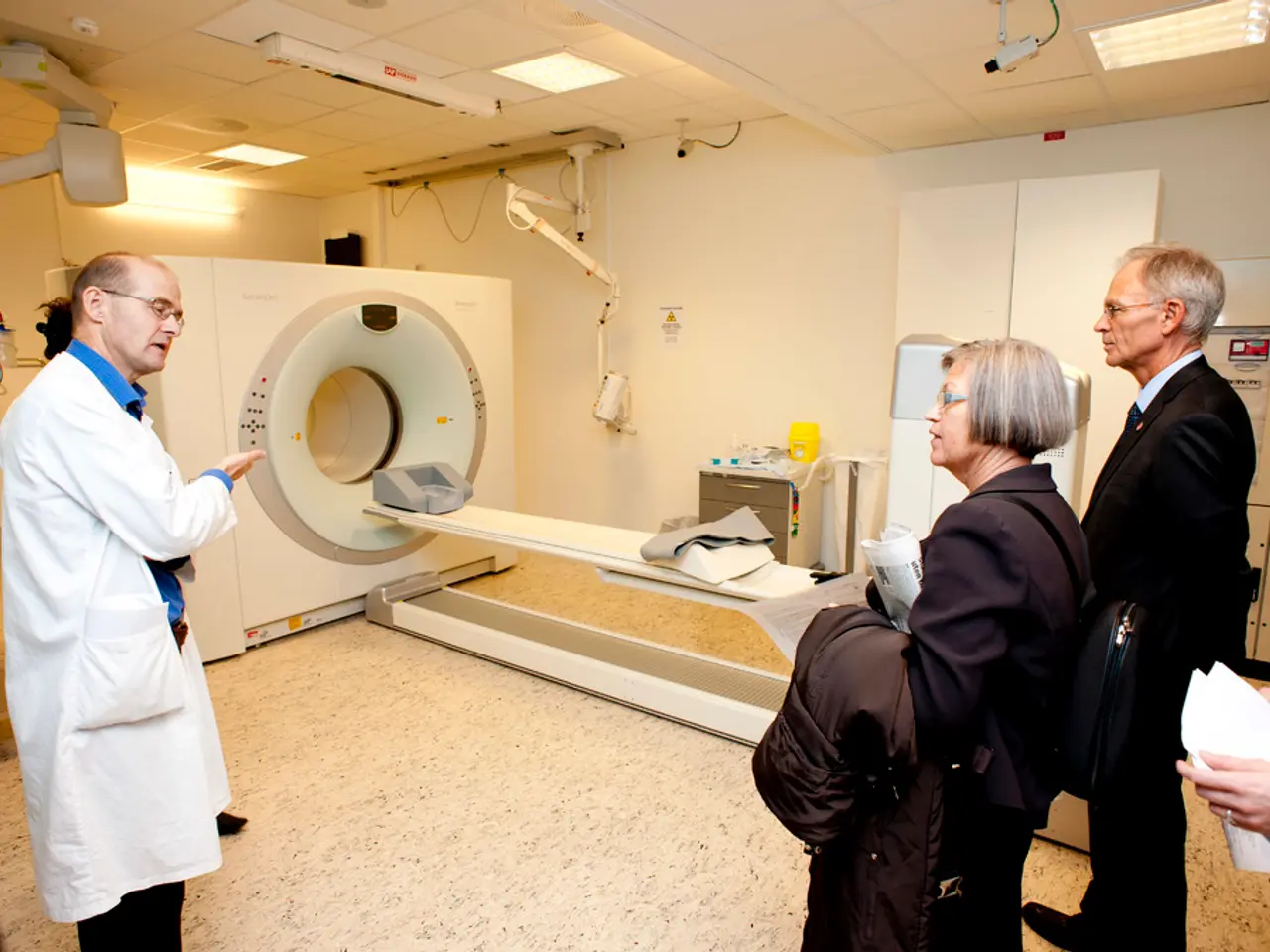
PET imaging, with its versatility in using various radiotracers tailored to different types of cancer and biological processes, is playing a crucial role. For instance, F-fluoromisonidazole (FMISO) is used to image hypoxia within tumors, while Ga-prostate-specific membrane antigen (PSMA) is particularly effective for detecting primary and metastatic prostate cancer. The most commonly used radiotracer in cancer imaging, F-FDG, allows PET to detect tumors that may not be visible on anatomic imaging, assess tumor activity, and evaluate treatment response.
Radiomics, a quantitative imaging data analysis method, is another powerful tool. It gathers quantitative information from medical images to build classification or predictive models for cancer diagnosis, prognosis, and prediction of treatment response. By incorporating PET data into radiomic analysis, radiomic features can be computed to reflect both the anatomic and functional characteristics of the tumor, providing a more detailed characterization of the tumor and potentially leading to more accurate cancer diagnosis, prognosis, and prediction of treatment response.
The synergy between PET imaging and radiomics is proving to be a potent combination. AI algorithms analyse these radiomic features to detect subtle patterns imperceptible to human observers, supporting early tumor detection, precise margin delineation, and prediction of treatment response. AI-driven radiomic analyses are surpassing conventional imaging interpretation.
Moreover, AI systems are being developed to integrate radiomic features from PET and CT with molecular and clinical data, such as genomics, enabling superior tumor characterization and prognostic stratification. This holistic approach is expected to improve cancer screening accuracy, reduce false positives/negatives, assist in surgical planning with better tumor boundary definition, and enable noninvasive monitoring of tumor evolution during therapy.
Future directions in this field include hybrid PET/MRI systems integrated with AI radiomics models combined with multi-omics data (genomics, transcriptomics, proteomics) for a more precise and reproducible diagnostic platform. This approach aims to enhance clinical utility and personalize treatment by correlating imaging phenotypes with molecular tumor signatures.
Emerging techniques like nanoparticle-based probes visible in PET and other imaging forms offer dual diagnostic and therapeutic ("theranostic") potential. This convergence may enable targeted drug delivery guided by real-time PET imaging and AI analysis for adaptive treatment.
Advanced AI models that integrate PET imaging features with genetic risk factors and clinical variables are expected to improve early cancer detection and better predict disease recurrence or treatment resistance, enabling tailored interventions and follow-up.
The future calls for tighter collaboration between academia, biotechnology, and pharmaceutical industries to refine imaging protocols, data interoperability, and AI algorithm transparency to ensure robust, ethical clinical deployment of these technologies.
In summary, the frontier of personalized cancer care lies in the convergence of PET imaging with AI-powered radiomics and multimodal data integration. This collective approach enhances tumor phenotyping, optimizes therapeutic decisions, and paves the way for precision oncology that adapts dynamically to individual patient tumor biology and treatment responses.
- The integration of Positron Emission Tomography (PET) imaging with artificial intelligence (AI) is transforming the diagnosis and treatment of cancer, particularly through the use of radiomics for more detailed characterization of tumors.
- AI algorithms, analyzing radiomic features from PET imaging, can detect subtle patterns imperceptible to human observers, supporting early tumor detection, precise margin delineation, and prediction of treatment response.
- Future advancements in personalized cancer care are expected to include hybrid PET/MRI systems integrated with AI radiomics models, multimodal data integration, and advanced AI models that improve early cancer detection by correlating imaging phenotypes with molecular tumor signatures.


